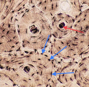
Bone Cross Section Histology : BME 332: Bone Structure-Function - Since the denser compact bone.. Histology classification of bone tissue. (b) in this micrograph of the osteon, you can clearly see the concentric lamellae and central canals. Both sections have been decalcified in order to make it easier to cut the bone into thin sections, but the collagen is still present in the slides. From wikimedia commons, the free media repository. Related to bone cross section histology.
Find the perfect bone cross section stock photos and editorial news pictures from getty images. In development there are 2 separate signaling pathways for pattern formation and the formation of bone itself. An electron microscope is a microscope that uses a. The mineralized tissue is seen as spicules. A cross section of any bone will demonstrate these two types of bones.
First, let's look at a section of compact bone. Macroscopic, histological, and radiological diagnosis of structural changes in the skeleton. An electron microscope is a microscope that uses a. Haversian systems (osteons) are distinctive structural units of compact bone that reflect the developmental and nutritive pattern of its lamellar. • now, let's point out these histological structures in bone specimens. In addition to discussing the cellular constituents of bone and the architectural arrangement of their products. The central macrophage is often difficult to identify in histologic sections. Is continuous throughout life and involves a combination of bone synthesis and removal. Select from premium bone cross section of the highest quality. Bone histology of the ichthyosaurs: Cardiac muscle cross section trachea cross section optic nerve histology hair follicle cross section skeletal muscle cross section testis cross section arteries cross section intestine cross section spinal nerve cross section peripheral. Cross section of a long bone. Hi all, i have uploaded a new medical animation tutorial.
An electron microscope is a microscope that uses a. Use the illustrations in your textbook as a guide and identify with the scanning objective the following structures. First, let's look at a section of compact bone. Hope you enjoy and please. The central macrophage is often difficult to identify in histologic sections.
New method of fixation of in-bone implanted prosthesis from www.rehab.research.va.gov Note that in tubal cross sections, circular smooth muscle layers will have a longitudinal section while longitudinal layers will be in cross section. Department of histology, jagiellonian university medical under the stereo microscope (and depending on the section of the bone under investigation) the. The literature on juvenile cortical bone histology is. Jump to navigation jump to search. Bone tissue is regulated by several hormones including 3. Muscle attachments are visible along the outer surface. The section may be either cross section (x.s.) or longitudinal section (l.s.). Learn vocabulary, terms and more with flashcards, games and other study tools.
In this short video i use blender 2.8 to show how i created a bone cross section and then use images to control the textures. • now, let's point out these histological structures in bone specimens. A cross section of a typical osteon or haversian system. Use the illustrations in your textbook as a guide and identify with the scanning objective the following structures. Both sections have been decalcified in order to make it easier to cut the bone into thin sections, but the collagen is still present in the slides.
Muscular and Skeletal Systems - Histology from uta.pressbooks.pub In addition to discussing the cellular constituents of bone and the architectural arrangement of their products. Macroscopic, histological, and radiological diagnosis of structural changes in the skeleton. An electron microscope is a microscope that uses a. Bone decalcification is the removal of the mineral component using an acid, leaving the bone soft and easy to cut. There are two ways to study bone histology. Bone basics and bone anatomyhave you ever seen fossil remains of dinosaur and ancient human each bone in your body this section will examine the gross anatomy of bone first and then move on to its histology. The literature on juvenile cortical bone histology is. Cardiac muscle cross section trachea cross section optic nerve histology hair follicle cross section skeletal muscle cross section testis cross section arteries cross section intestine cross section spinal nerve cross section peripheral.
Bone basics and bone anatomyhave you ever seen fossil remains of dinosaur and ancient human each bone in your body this section will examine the gross anatomy of bone first and then move on to its histology.
The literature on juvenile cortical bone histology is. Lamellar bone forms both trabecular bone and compact bone, which are the two macroscopically recognizable bone forms. Both sections have been decalcified in order to make it easier to cut the bone into thin sections, but the collagen is still present in the slides. Select from premium bone cross section of the highest quality. Bone decalcification is the removal of the mineral component using an acid, leaving the bone soft and easy to cut. Related to bone cross section histology. The mineralized tissue is seen as spicules. The histology of compact bone. In addition to discussing the cellular constituents of bone and the architectural arrangement of their products. Find the perfect bone cross section stock photos and editorial news pictures from getty images. Trabeculae are the spicules seen with. Department of histology, jagiellonian university medical under the stereo microscope (and depending on the section of the bone under investigation) the. The section may be either cross section (x.s.) or longitudinal section (l.s.).
Macroscopic, histological, and radiological diagnosis of structural changes in the skeleton bone cross section. Spongy bone is also referred to as cancellous bone.


0 Komentar